7 Riverview Drive, Guelph, Ontario, N1E 3R6,
Canada
Mon: 10:00am - 7:00 pm
Tu/W: 8:00am - 5:00 pm
Thur: 9:00am - 4:00 pm
Fri - Sat 9:00 am - 3:00 pm (alternating)

20-year-old front crown with underlying decay replaced with a custom shade-matched crown - A cosmetic challenge.
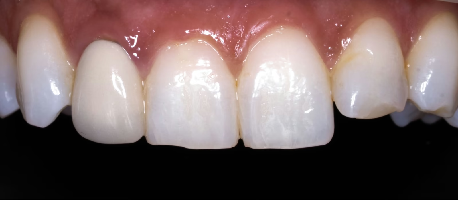
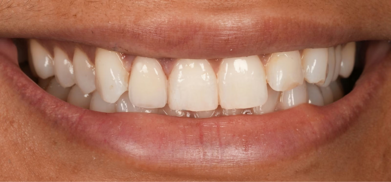
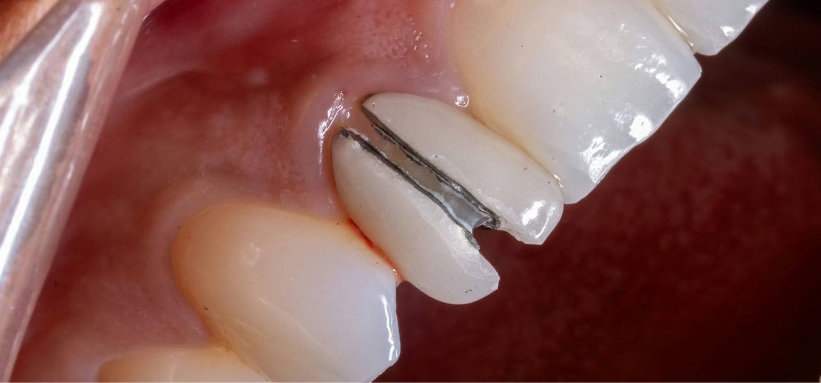
An upper lateral incisor with a porcelain-fused-to-metal crown which is over twenty years old. The existing crown does not match the patients dentition, is opaque, bulky, and has developed recurrent decay (a cavity) along the margin and has broken down over the years due to the patients history of bruxism (grinding) and clenching.
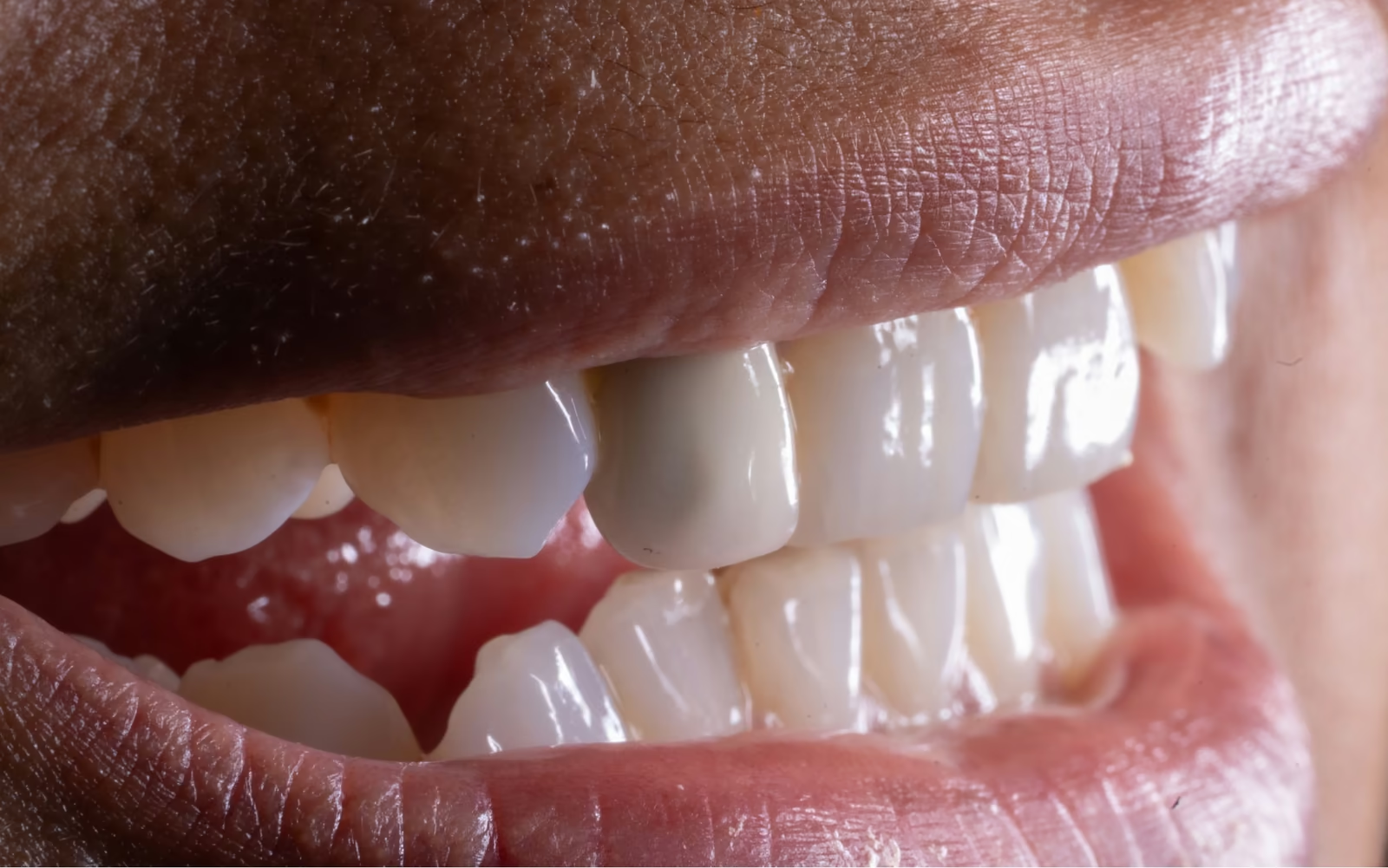
Cosmetically the old crown had held up reasonably for its age, but it had become identifiable: more opaque than the teeth around it, slightly off in shade, smooth where natural enamel has fine texture, and without translucency at the biting edge. Shade matching a single tooth crown to the adjacent dentition is among one of the most challenging cases in restorative dentistry.
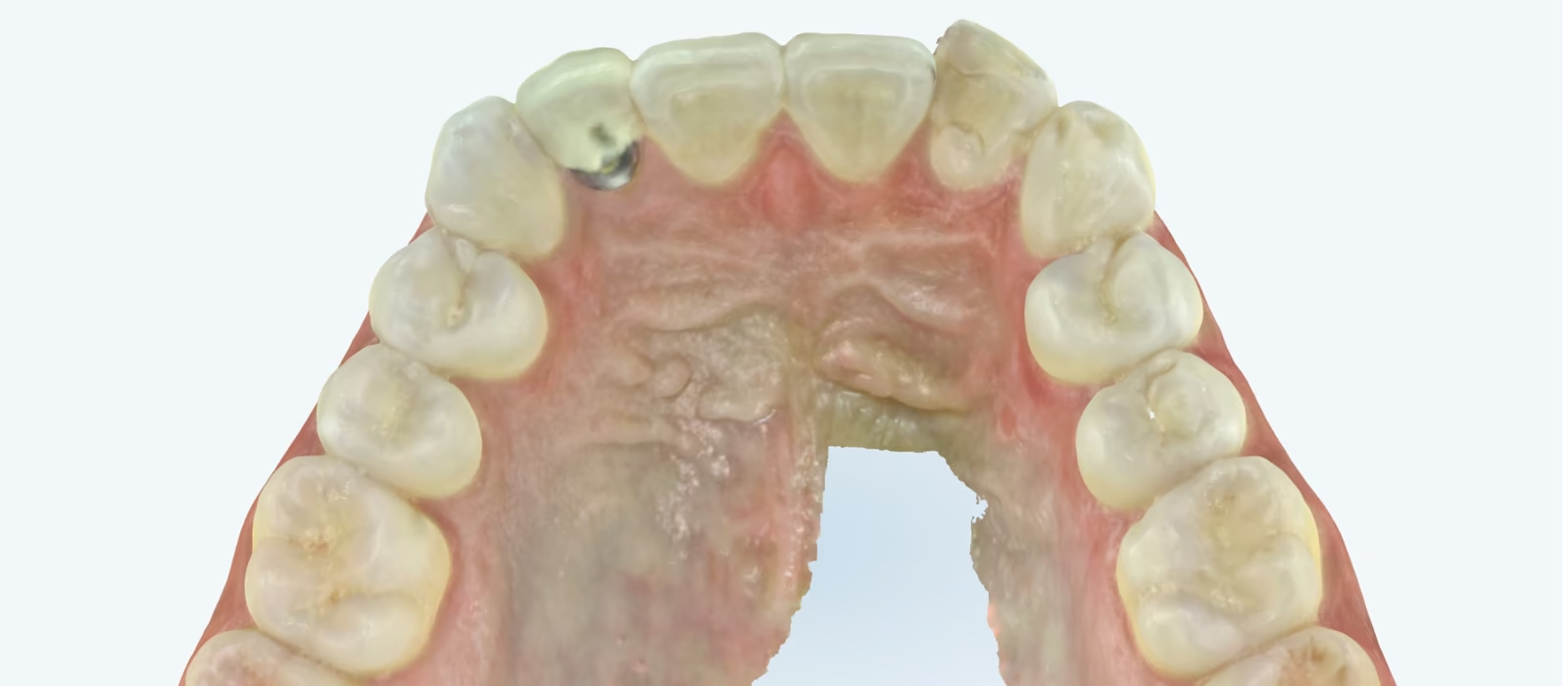
First we need to assess how deep the decay is. How deep it went could not be established while the crown was in place, because crown material blocks the X-ray view of the tooth beneath. Assessment therefore ran in two stages: radiograph, nerve vitality testing, percussion and palpation beforehand. This gives us an idea of the nerve status. Then crown removal, direct inspection once the crown was off and treatment of the decay.
The decay proved to be 1 to 2 mm deep, within dentin and clear of the nerve, which tested healthy. That meant a straightforward crown replacement, with no root canal needed.
The old crown was sectioned under magnification and lifted off without removing additional natural tooth. The preparation was refined, with the decayed area cleaned out and incorporated into the shape the new crown would fit over.
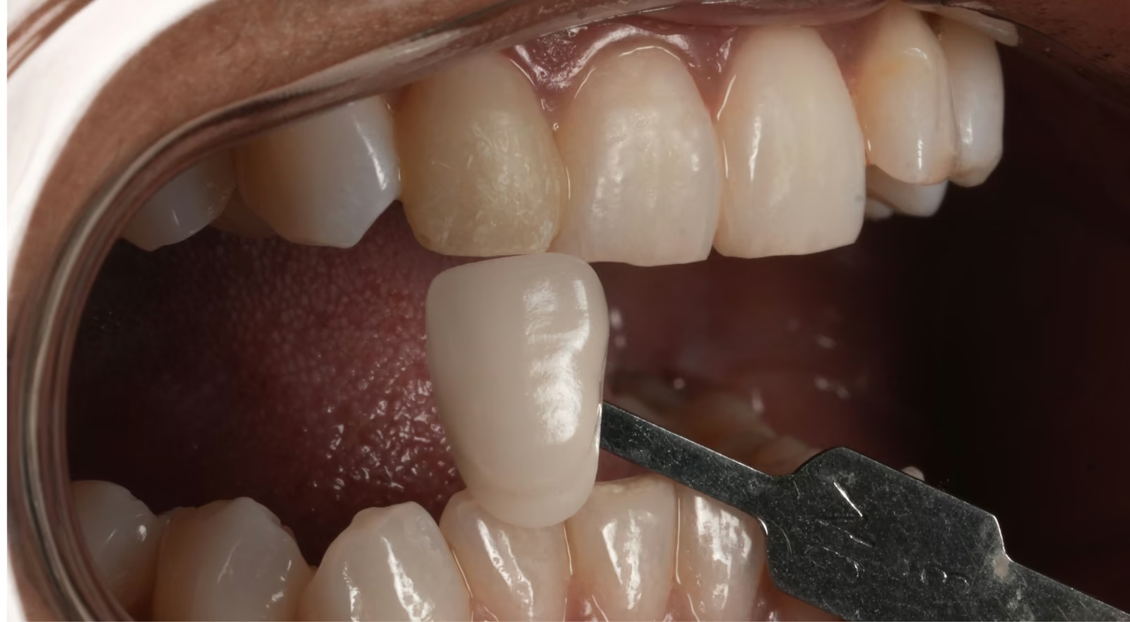
A digital scan was taken and a temporary crown fitted. The new crown was then shade-matched to the central incisor beside it — the most demanding part of the case. Around twenty to thirty calibrated photographs were taken with shade guides under different lighting, including cross-polarized images that strip surface glare so internal colour can be recorded, and a shade map was drawn up for the laboratory.
Zirconia was chosen deliberately: the prepared tooth underneath was dark, and zirconia is opaque enough to mask it where a more translucent material would have let the darkness show through. The facial surface was cut back and layered in porcelain for genuine translucency.
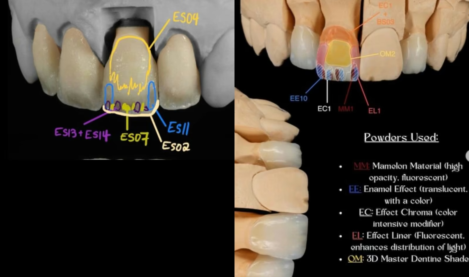
The final crown is then manually painted and layered with varying kinds of porcelain, this requires a considerable degree of artistry, texture is recreated and then polished.
The crown was tried in and assessed under several lighting conditions, with the patient evaluating it in natural light before it was cemented. The bite was balanced at delivery.
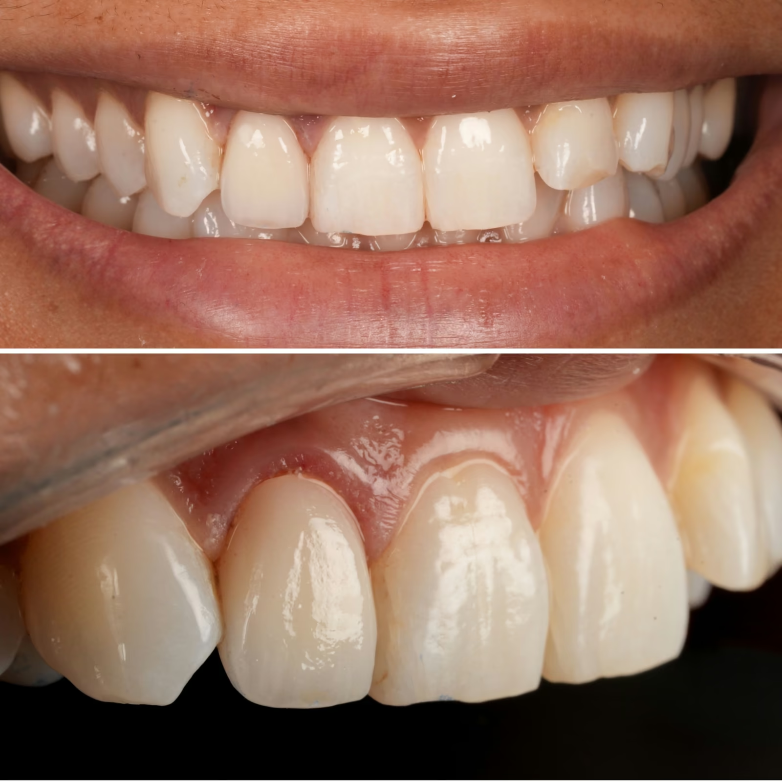
This image shows the crown immediately after cementation.
The tissue then heals and appears more natural around the crown as the weeks pass by. A night guard was made. The previous crown broke down under grinding forces, and replacing it without addressing that force would have left the same problem in place. Worth knowing: a night guard protects teeth while worn but does not stop clenching, and it is not a treatment for sleep-disordered breathing, that is a separate medical conversation.
Individual results vary; matching a single crown to natural teeth beside it is among the harder tasks in dentistry, and what is achievable depends on the tooth and its neighbours. This case is not meant to provide medical advice but documents a clinical case performed at sunny dental centre. Individuals should refer to their dentist or medical providers for individual or general medical/dental advice.
Sunny Dental Centre is a general dental practice. Dr. Mandip Puri, DDS, is registered with the Royal College of Dental Surgeons of Ontario as a general practitioner, not a specialist.
Was freezing needed?
Yes. Crown removal and preparation ordinarily require local anesthetic.
Was there pain afterwards?
No post-operative pain was reported in this case, which is common after crown replacement. Pressure discomfort can occur when the bite is not perfectly balanced and settles within a few days or after a small adjustment.
Could the decay just be filled instead?
Sometimes, if it is small and accessible at the edge. More often decay tracks underneath where it cannot be seen or fully removed. Here the crown also had structural breakdown on the tongue side, which settled the question.
Why did the old crown look darker than the other teeth?
Usually one of three things: it was matched to teeth that have since changed colour, the material lacks the translucency of enamel, or a metal substructure is showing at the gumline as tissue recedes. Whitening does not change a crown, which is why whitening is planned before crown work rather than after.
For more details refer to the blog post which reviews this case in detail.
Individual results vary. This case illustrates work completed at Sunny Dental Centre and is shared with written patient consent. What is achievable in your own case depends on your teeth, your bite and your goals. Sunny Dental Centre is a general dental practice; Dr. Mandip Puri, DDS, is registered with the Royal College of Dental Surgeons of Ontario as a general practitioner, not a specialist.
Every case is different. Book a consultation and we will tell you what is realistic for your teeth.
7 Riverview Drive, Guelph, Ontario, N1E 3R6,
Canada
Mon: 10:00am - 7:00 pm
Tu/W: 8:00am - 5:00 pm
Thur: 9:00am - 4:00 pm
Fri - Sat 9:00 am - 3:00 pm (alternating)